Retina Surgery
Wolfe Eye Clinic has expertise in all modern retina surgery techniques. The most common retinal procedures performed are vitrectomy and scleral buckle surgery. Retina surgery is performed in a special operating room that has high tech equipment and expert staff trained for retina procedures. When possible, we always try and avoid surgery. However, in cases where medical treatments or other options are not possible, you should be assured that our fellowship trained retina surgeons are experts in handling diseases that require surgical intervention. Wolfe Eye Clinic retina specialists perform retina surgeries at trusted hospital locations in Cedar Rapids and Cedar Falls. In addition, Wolfe Eye Clinic retina surgeons perform many Iowa retina surgeries at our state-of-the-art Wolfe Surgery Center in Des Moines, which is complete with some of the most advanced ophthalmic surgical technology available today.
Vitrectomy Surgery for Retina Conditions
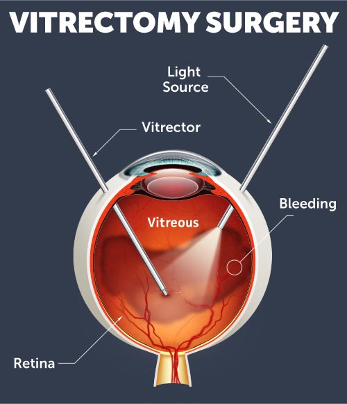
 A vitrectomy is a type of eye surgery that treats various problems with the retina and vitreous. Vitrectomy is the surgical removal of the vitreous gel from the large chamber of the eye. Common reasons for vitrectomy surgery include:
-
Repairing macular holes
-
Removing blood or floaters keeping light from focusing properly on the retina
-
Removing abnormal traction on the surface of the retina and causing changes in vision (epiretinal membranes, vitreomacular adhesion)
-
Repairing a detached retina
-
Removing a foreign object inside the eye from an injury
Preparing for Vitrectomy Surgery
In preparation for the vitrectomy surgery, you are sedated, and the eye is anesthetized (numbed) so you are comfortable during the procedure. The eye is prepared with antiseptic solution and a sterile drape is applied. An eyelid speculum is used to keep the eyelid open. The other eye is covered for protection and patients generally close their non-operative eye and are able to rest during the surgery.
About the Vitrectomy Surgery Procedure
During a vitrectomy procedure, the retina surgeon removes the vitreous gel from your eye through very small trocars, tiny ports that are placed in the pars plana, a special part of the eye where the vitreous can be safely removed. Once the vitreous is removed then additional procedures are performed depending on the reason for surgery. This could include removing epiretinal membranes, performing laser, or reattaching the retina. For more information about vitrectomy surgery, please see our pages with information for specific conditions. At the end of surgery, the vitreous is replaced with either a special salt solution, gas, or silicone oil depending on the reason for surgery. The vitreous gel is no longer needed for proper function and eventually the eye replaces the gas or salt solution with its own natural fluid. The tiny ports placed at the beginning of surgery are removed and the eye is patched. In many cases, stitches are not needed to close the eye. There is usually minimal pain or discomfort used after surgery. If you have a gas bubble in your eye you may need to position yourself in a certain way and avoid changing elevations or flying in an airplane. Most patients do quite well after vitrectomy surgery. In many cases, patients can resume all of their normal activities shortly after surgery. It is important to understand that if you have your own natural lens in the eye, it will become cloudy and develop a cataract more rapidly than before the vitrectomy. Your eye doctor will guide you if cataract surgery becomes necessary.
Recovering From Vitrectomy Surgery
Many people ask, “Is vitrectomy surgery a major surgery”? While any surgery performed on the eye is serious, most vitrectomy surgeries are performed in an  outpatient setting and are comfortable. Our retina surgeons use the latest microsurgical techniques and stitches are not needed for most patients making this retina surgery recovery process quick. Discomfort after surgery is minor and you can resume many of your normal activities right after surgery. The goals of vitrectomy surgery are to treat an abnormal condition causing vision loss, to reduce the likelihood of recurrence and to minimize the risk of complications. Complications of vitrectomy surgery are rare, but could include infection, bleeding, high or low eye pressure, cataract, retinal detachment, and loss of vision. It is important that you follow the instructions given by your retina doctor post-surgery and attend any follow-up appointments with your retina specialist.
Scleral Buckle Surgery for Retina Detachment
Scleral buckling surgery is a common way to treat a retinal detachment. It is a method of closing retinal breaks and supporting a tear, hole or break in the retina that has caused the retinal detachment. A scleral buckle is a piece of silicone sponge, rubber, or semi-hard plastic. It is placed on the sclera, or outside white layer of the eye, underneath the skin and the muscles of the eye. Your doctor will discuss the options for repairing your detached retina and let you know if a scleral buckle surgery is right for you. In some cases, a scleral buckle can be combined with a vitrectomy.
About the Scleral Buckle Procedure
This procedure is performed in a special operating room under sedation or while you are asleep. The eye is prepared with antiseptic solution, sterile drapes are placed to keep everything clean, and an eyelid holder is used to gently hold the eyelids in place. Small incisions are made in the clear skin (conjunctiva) covering the white part of the eye. The tears or holes in the retina that caused the detachment are located and treated with freezing therapy or laser. A special silicone band (scleral buckle) is placed under the skin and muscles of the eye and around the globe, gently pushing on the wall of the eye to keep the retina attached. Once the fluid has been drained from under the retina, the skin of the eye is closed with dissolving stitches. In some cases, your doctor may inject a gas bubble to help hold the retina in place from the inside. Your eye will be patched and then you will be able to go home to rest and relax. You might have to hold your head in a certain position after surgery to put the bubble up against the right part of the retina.
Recovering from Scleral Buckle Surgery
After the procedure, you may have some discomfort for a few days and your vision may be blurry. It is common for the eyelid to be swollen with bruising and the eye to be red. Most patients are comfortable with over the counter pain medications. You will take some eye drops to prevent infection and promote healing. It is important to follow close instructions given by your retina surgeon and contact a retina doctor right away if you have concerns or notice any changes such as increasing redness, pain or swelling. Experiencing any new floaters, flashes of light or changes in your field of vision during retina surgery recovery would also be a warning sign to call your doctor.
Contact Iowa's Retina Surgeons Near You
Wolfe Eye Clinic offers retina surgery in Iowa at locations throughout the state, including the Cedar Rapids, Cedar Falls and Des Moines areas making the search for retina surgeons near you easy. If you have any retina-related questions or would like to schedule an appointment with a retina specialist, please call us at (833) 474-5850 or request information here.