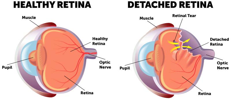
What is a retinal tear and what is a retinal detachment?
The retina is the light-sensitive layer of tissue that lines the inside of the eye and sends visual messages through the optic nerve to the brain. The larger back chamber of the eye is filled with a clear gel called the vitreous. As a normal part of the aging process, the vitreous gel separates from the retina. You can develop a retina tear when this gel pulls on the fragile retina. Retina tears can cause bleeding in the eye that can blur your vision or create floaters. The main problem with a retina tear is that it can lead to a detached retina, a sight-threatening problem.
Learn more details about a detached retina, what you can expect and how it may be treated.
How does the retina detach?
Once the retina develops a tear or hole, fluid from within the eye can move through this opening and collect under the retina, detaching it from the wall of the eye. The retina receives its oxygen and nutrition from a collection of blood vessels between the retina and the wall of the eye. When the retina becomes detached it becomes sick resulting in vision loss.
What causes retinal tears and detachment?
Many patients ask, what causes retinal tears? Or how does a detached retina happen? Almost all retina detachments are caused by vitreous separation as a result of normal aging. Retina tears or detachments caused by trauma and other diseases are rare but can occur. If you are very near sighted (high myopia) you are at an increased risk of having a tear or detachment. Some people have weak areas in their retina that can also increase the chance of having the retina tear with vitreous separation. In certain cases, these areas can be treated to decrease the change of problems.
If you have a torn or detached retina in one eye you are at a higher risk of developing it in your other eye. In almost all cases of a torn retina or detachment there is nothing that you did to cause the problem.

What are symptoms of a retina tear or detachment?
How do you know if you have a retinal tear or detachment? There are a few signs you can look for to help determine if you should schedule an appointment with you eye doctor. These include:
- Sudden appearance of many floaters (small specks or “cobwebs”)
- Blurry vision
- Flashes of light in one or both eyes
- A curtain-like shadow in your visual field
If you have any of these symptoms, please call your eye care doctor immediately.
Your eye doctor will perform a dilated examination of the eyes to closely inspect the retina. Retina tears and detachments are typically painless. Retinal detachment repair has a much better vision outcome if it is performed before the center part of the vision is involved. Responding to the warning signs quickly may help prevent permanent vision loss.
Learn more about the signs and symptoms of a retina tear and the steps you should take to protect your vision.
Treatment for Retinal Tears and Detachment
Once a diagnosis of torn or detached retina is made you will need to see a retina specialist who will perform a retinal examination that includes an instrument with a bright light and special lenses to examine the back of your eye. This type of specialized device provides a highly detailed view of your whole eye, allowing them to examine your retina for any retinal holes, tears or detachments. Sometimes photographs are taken to assist in making a diagnosis or documenting your retina problems.
Prevent Retinal Detachment with Retinal Tear Treatment
Retinal tears can be treated to prevent retinal detachment. This treatment at Wolfe Eye Clinic is almost always performed in the comfort of our retina clinic locations throughout Iowa. Your retina specialist can use a special laser to surround the tear and seal it to prevent it from detaching. A special lens is used to focus the laser, a concentrated light, on the retina to deliver small burns or “welds.” Over time these laser spots heal to hold the retina in place. After the laser, you will feel like a bright light was shined in the eye. This feeling returns to normal over a few minutes to hours. Laser treatment can be repeated as necessary.
In some cases, retina tears are frozen in a procedure in the office called cryopexy. After the eye is made comfortable with a numbing treatment, a special freezing probe is directed under the tear and the freezing treatment delivered around the tear. After treatment the eye forms an adhesion holding the retina in place.
Recovering from Retina Tear Treatment
 Most people recover well after a treatment for a retinal tear. Patients are able to return home to relax in the comfort of their own homes. Most eyes will feel comfortable but dazed as if a they stared at a very bright light. These symptoms, including the dilation, should wear off over the rest of the day. Most find themselves able to go about their normal daily routines shortly after the procedure. Typically, patients can return to full activity the very next day.
In some rare cases, a retinal detachment will happen despite excellent laser treatment. This usually occurs from new tears or the development of strong traction on the retina breaking through the treatment. Your retina specialist will monitor you carefully after treatment to make sure you respond as planned.
Retinal Detachment Repair
Timing can be very important for repair of a retinal detachment. Your retina specialist will try and fix the detachment before the center part of the retina, known as the macula, is impacted. If your macula is not yet involved, it will be important to try and fix the retina urgently.
If the center part of the retina is already involved, the repair will usually occur sometime within a couple of weeks. There are several different methods to repair a detached retina. Which method you and your retina doctor select depends on your specific condition. There are a few options for retinal detachment surgery, and your retina specialist may even use a combination of approaches to try and restore the retina to health and improve your vision.
Can a detached retina heal on its own?
Very rarely, retinal detachments are not noticed by the patient and can heal on their own. The vast majority of retinal detachments progress to irreversible vision loss if left untreated so it is important to monitor any changes noticed in your vision.
Vitrectomy Surgery for Retina Repair
A vitrectomy is performed in a special operating room for retina surgery, usually under sedation. At Wolfe Eye Clinic this is generally performed at our Wolfe Surgery Center in Des Moines but can also be performed with our trusted hospital locations around the state by a Wolfe Eye Clinic retina specialist. After you are comfortable, the retina surgeon removes the vitreous gel using small instruments through very small holes in a special part of the white part of the eye (sclera).
The retina is flattened and then laser is applied. Next, a gas bubble is placed in the large chamber of the eye to hold the retina in place as the eye heals. In some instances, particularly if your retina has scar tissue that has formed, silicone oil is placed in the eye to hold the retina in place. In many cases no stitches are needed. The eye is then patched with a bandage and you are instructed by your retina specialist to go home and relax.
Recovering from Vitrectomy Surgery
After a vitrectomy, you may need to hold your head in a certain position depending on the locations of your retina tears. Your activity is usually restricted after surgery but there is usually little or no discomfort after surgery. You will take some eye drops to prevent infection and promote healing. During the healing process the gas bubble shrinks and your eye replaces it with its own natural fluid. Vision improves as the gas bubble shrinks. While you have a gas bubble inside the eye it is important to stay off your back, not fly in an airplane or change altitudes, and inform your doctors if you need any other type of surgery.
Learn More About Vitrectomy Surgery
Scleral Buckle Surgery for Retina Detachments
Retina detachments can be repaired with scleral buckle surgery. This procedure is performed in a special operating room under sedation or while you are asleep. The tears or holes in the retina that caused the detachment are located and treated with freezing therapy or laser. A special silicone band (scleral buckle) is placed around the eye, under the skin and muscles, gently pushing on the wall of the eye to keep the retina attached. Once the fluid has been drained from under the retina, the skin of the eye closed with dissolving stitches. In some cases, your doctor may inject a gas bubble to help hold the retina in place from the inside. Your eye will be patched and then you will be able to go home to rest and relax. You might have to hold your head in a certain position after surgery if the bubble is also used. The eye can experience some discomfort after surgery that is usually treated with over the counter pain medications. You will take some eye drops to prevent infection and promote healing.
Pneumatic Retinopexy for Retina Detachments
Some types of retina detachments can be repaired with an in-office procedure called a pneumatic retinopexy. During this procedure, after the eye is numbed, the retina tears are treated with freezing or laser and a gas bubble is injected. The gas bubble floats and pushes the retina back into place. After the procedure you are able to go home and relax. You will need to keep your head in a certain position to hold the retina in place as things heal. You might take some eye drops to prevent infection and promote healing.
What is the recovery like after retinal detachment repair?
Many aspects of your detached retina surgery recovery time will be determined by the type of retinal detachment and the procedure needed to fix it. Your retina surgeon will review this in detail as this is specific to your situation. One big factor in your detached retina surgery recovery time is your gas bubble and the positioning required to keep the retina in place while you heal. There are two types of gas that are used. One lasts 2 weeks and the other lasts 2 months. Your surgeon will select the proper gas bubble for your procedure, always trying to use the bubble that dissolves faster. If you have silicone oil placed in the eye, it is usually removed several months after the eye has healed.
A majority of retina detachments are able to be fixed with a single procedure. There are some cases where the retina becomes detached again, usually from the development of scar tissue. If the retina becomes detached again, additional surgery will be necessary. The vision is often quite blurry right after your procedure. This gets better as the eye heals. You will be monitored very closely as your retina heals. It is important to monitor your vision closely and report changes to your retina surgeon immediately. Your retina doctor will advise you as to how much physical activity you can pursue immediately after the procedure. You should also keep a lookout for a significant increase in floaters, flashes or loss of peripheral vision. Wolfe Eye Clinic has doctors on call 24-hours a day to address any emergencies that arise.
How do you sleep after a retinal detachment surgery?
If a gas bubble is used to fix your retinal detachment, staying off your back during sleep is very important. Sleeping on your stomach or sides is usually allowed but rest assured that your retina surgeon will instruct you on this according to your situation.
Can you drive after retinal detachment surgery?
Driving is often limited after surgery due to the blurry vision caused by the gas bubble or healing process. It is important to exercise caution and consult your retina specialist with any questions regarding the healing process.
Visual Results from Retinal Detachment Repair
The visual outcome is not always predictable after retinal detachment repair. The final visual result may not be known for up to several months following surgery. Even after successful repair, the vision often will not return to normal. The biggest factor in having good vision after surgery is whether the center part of your retina, the macula, was detached before it was repaired. Visual results are best if the retinal detachment is repaired before the macula, or center region of the retina, detaches. When the retina is detached it is separated from crucial nutrients and oxygen supply causing permanent damage can occur to the retina. While surgery can attach the retina, it cannot reverse that damage that was done to the retina. It is very common to have blurred or distorted vision. These vision problems cannot usually be corrected with glasses, contact lenses, medications or other types of surgery.
It is important to monitor your vision closely and report changes to your retina surgeon immediately. Your retina doctor will advise you as to how much physical activity you can pursue immediately after the procedure. You should also keep a lookout for a significant increase in floaters, flashes or loss of peripheral vision. Wolfe Eye Clinic has doctors on call 24-hours a day to address any emergencies that arise.
Find a Detached Retina Surgeon Near You
Wolfe Eye Clinic has many retina specialists in Iowa and they have expertise in treating detached retinas and many other conditions of the eye. Wolfe Eye Clinic offers complete retina disease services in Iowa, including Ames, Ankeny, Cedar Falls, Cedar Rapids, Des Moines, Fort Dodge, Iowa City, Marshalltown, Ottumwa, Spencer, Waterloo, and Pleasant Hill. You can request more information on retina conditions here, or give us a call at (833) 474-5850.